
ATG3 Primary Antibody
Item Information
Catalog #
Size
Price
Description
This gene encodes a ubiquitin-like-conjugating enzyme and is a component of ubiquitination-like systems involved in autophagy, the process of degradation, turnover and recycling of cytoplasmic constituents in eukaryotic cells. This protein is known to play a role in regulation of autophagy during cell death. A pseudogene of this gene is located on chromosome 20. Alternative splicing results in multiple transcript variants encoding different isoforms.
Product Overview
Entrez GenelD
64422
Aliases
APG3; APG3L; PC3-96; APG3-LIKE
Clone#
7A1D1
Host / Isotype
Mouse / IgG1
Species Reactivity
Human, Monkey
Immunogen
Purified recombinant fragment of human ATG3 (AA: 1-100) expressed in E. Coli.
Formulation
Purified antibody in PBS with 0.05% sodium azide
Storage
Store at 4°C short term. Aliquot and store at -20°C long term. Avoid freeze/thaw cycles.
Product Applications
WB (Western Blot)
1/500 - 1/2000
IHC_P(Immunohistochemistry)
1/200 - 1/1000
ICC (Immunocytochemistry)
1/200 - 1/1000
ELISA
1/10000
References
1.Mol Biol Rep. 2014;41(4):2093-9.
2.Apoptosis. 2012 Aug;17(8):810-20.
2.Apoptosis. 2012 Aug;17(8):810-20.
Product Image
Elisa
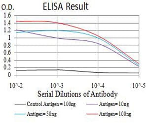
Figure 1: Black line: Control Antigen (100 ng);Purple line: Antigen (10ng); Blue line: Antigen (50 ng); Red line:Antigen (100 ng)
Western Blot
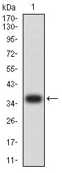
Figure 2:Western blot analysis using ATG3 mAb against human ATG3 (AA: 1-100) recombinant protein. (Expected MW is 37.3 kDa)
Western Blot
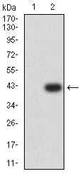
Figure 3:Western blot analysis using ATG3 mAb against HEK293 (1) and ATG3 (AA: 1-100)-hIgGFc transfected HEK293 (2) cell lysate.
Western Blot
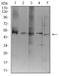
Figure 4:Western blot analysis using ATG3 mouse mAb against Jurkat (1), K562 (2), Hela (3), THP-1 (4), and COS7 (5) cell lysate.
Immunofluorescence analysis
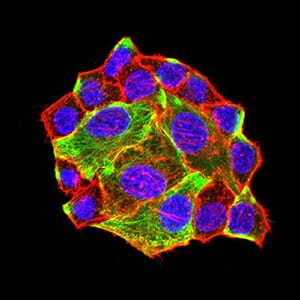
Figure 5:Immunofluorescence analysis of Hela cells using ATG3 mouse mAb (green). Blue: DRAQ5 fluorescent DNA dye. Red: Actin filaments have been labeled with Alexa Fluor- 555 phalloidin. Secondary antibody from Fisher (Cat#: 35503)
Immunofluorescence analysis
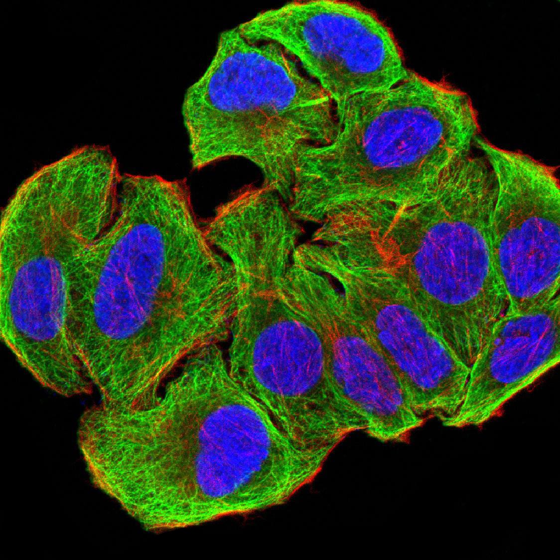
Figure 6:Immunofluorescence analysis of SMMC-7721 cells using ATG3 mouse mAb (green). Blue: DRAQ5 fluorescent DNA dye. Red: Actin filaments have been labeled with Alexa Fluor- 555 phalloidin. Secondary antibody from Fisher (Cat#: 35503)
Immunohistochemical analysis
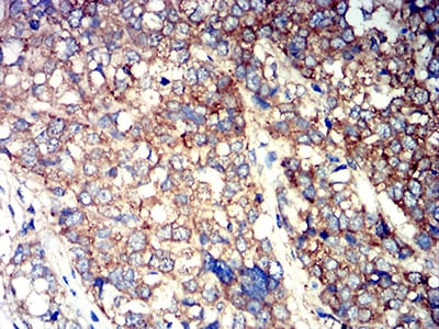
Figure 7:Immunohistochemical analysis of paraffin-embedded breast cancer tissues using ATG3 mouse mAb with DAB staining.
Immunohistochemical analysis
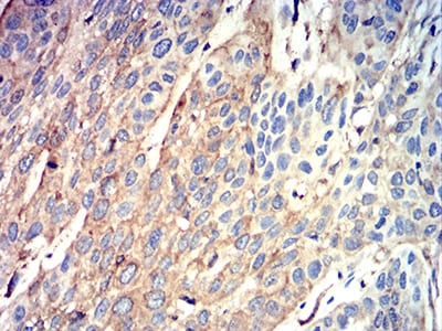
Figure 8:Immunohistochemical analysis of paraffin-embedded esophageal cancer tissues using ATG3 mouse mAb with DAB staining.
For Research Use Only. Not for use in diagnostic procedures.