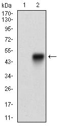
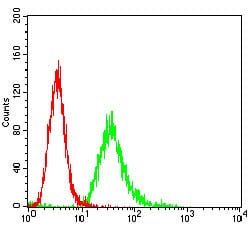
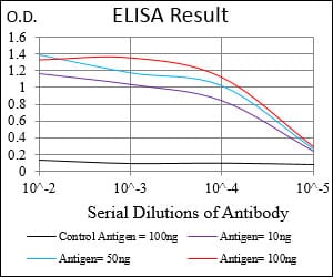
ANPEP Primary Antibody

Item Information
Catalog #
Size
Price
Description

Aminopeptidase N is located in the small-intestinal and renal microvillar membrane, and also in other plasma membranes. In the small intestine aminopeptidase N plays a role in the final digestion of peptides generated from hydrolysis of proteins by gastric and pancreatic proteases. Its function in proximal tubular epithelial cells and other cell types is less clear. The large extracellular carboxyterminal domain contains a pentapeptide consensus sequence characteristic of members of the zinc-binding metalloproteinase superfamily. Sequence comparisons with known enzymes of this class showed that CD13 and aminopeptidase N are identical. The latter enzyme was thought to be involved in the metabolism of regulatory peptides by diverse cell types, including small intestinal and renal tubular epithelial cells, macrophages, granulocytes, and synaptic membranes from the CNS. Human aminopeptidase N is a receptor for one strain of human coronavirus that is an important cause of upper respiratory tract infections. Defects in this gene appear to be a cause of various types of leukemia or lymphoma.

Product Overview
Entrez GenelD
290
Aliases
APN; CD13; LAP1; P150; PEPN; GP150
Clone#
1C7D7
Host / Isotype
Mouse / IgG1
Species Reactivity
Human
Immunogen
Purified recombinant fragment of human ANPEP (AA: Extra(781-967)) expressed in E. Coli.
Formulation
Purified antibody from tissue culture in PBS with 0.05% sodium azide
Storage
4°C; -20°C for long term storage
Product Applications
WB (Western Blot)
1/500 - 1/2000
FCM (Flow Cytometry)
1/200 - 1/400
ELISA
1/10000
References
1. Br J Cancer. 2013 Feb 5;108(2):420-8.
2. Ann Surg Oncol. 2012 Jul;19 Suppl 3:S539-48.
Product Image
Western Blot
Figure 1: Western blot analysis using ANPEP mAb against human ANPEP (AA: Extra(781-967)) recombinant protein. (Expected MW is 47.4 kDa)
Western Blot
Figure 2: Western blot analysis using ANPEP mAb against HEK293 (1) and ANPEP (AA: Extra(781-967))-hIgGFc transfected HEK293 (2) cell lysate.
Flow cytometric
Figure 3: Flow cytometric analysis of Hela cells using ANPEP mouse mAb (green) and negative control (red).
Elisa
Black line: Control Antigen (100 ng); Purple line: Antigen(10ng); Blue line: Antigen (50 ng); Red line: Antigen (100 ng);
For Research Use Only. Not for use in diagnostic procedures.