
ALCAM Primary Antibody
Item Information
Catalog #
Size
Price
Description
This gene encodes activated leukocyte cell adhesion molecule (ALCAM), also known as CD166 (cluster of differentiation 166), which is a member of a subfamily of immunoglobulin receptors with five immunoglobulin-like domains (VVC2C2C2) in the extracellular domain. This protein binds to T-cell differentiation antigene CD6, and is implicated in the processes of cell adhesion and migration. Multiple alternatively spliced transcript variants encoding different isoforms have been found.
Product Overview
Entrez GenelD
214
Aliases
MEMD; CD166; FLJ38514; MGC71733
Clone#
4H9A5
Host / Isotype
Mouse / IgG2b
Species Reactivity
Human, Mouse
Immunogen
Purified recombinant fragment of human ALCAM (AA: 48-216) expressed in E. Coli.
Formulation
Purified antibody in PBS with 0.05% sodium azide
Storage
Store at 4°C short term. Aliquot and store at -20°C long term. Avoid freeze/thaw cycles.
Product Applications
WB (Western Blot)
1/500 - 1/2000
IHC_P(Immunohistochemistry)
1/200 - 1/1000
FCM (Flow Cytometry)
1/200 - 1/400
ELISA
1/10000
References
1. Vascul Pharmacol. 2011 Mar-Jun;54(3-6):93-9.
2. Int J Gynecol Cancer. 2011 Apr;21(3):523-8.
2. Int J Gynecol Cancer. 2011 Apr;21(3):523-8.
Product Image
Western Blot

Figure 1: Western blot analysis using ALCAM mAb against human ALCAM recombinant protein. (Expected MW is 44.9 kDa)
Western Blot

Figure 2: Western blot analysis using ALCAM mAb against HEK293 (1) and ALCAM (AA: 48-216)-hIgGFc transfected HEK293 (2) cell lysate.
Western Blot

Figure 3: Western blot analysis using ALCAM mouse mAb against NIH/3T3 cell lysate.
Flow cytometric

Figure 4: Flow cytometric analysis of Jurkat cells using ALCAM mouse mAb (green) and negative control (red).
Immunohistochemical analysis

Figure 5: Immunohistochemical analysis of paraffin-embedded prostate cancer tissues using ALCAM mouse mAb with DAB staining.
Immunohistochemical analysis

Figure 6: Immunohistochemical analysis of paraffin-embedded bladder cancer tissues using ALCAM mouse mAb with DAB staining.
Elisa
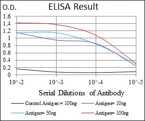
Black line: Control Antigen (100 ng); Purple line: Antigen(10ng); Blue line: Antigen (50 ng); Red line: Antigen (100 ng);
For Research Use Only. Not for use in diagnostic procedures.