
ACTA2 Primary Antibody
Item Information
Catalog #
Size
Price
Description
The protein encoded by this gene belongs to the actin family of proteins, which are highly conserved proteins that play a role in cell motility, structure and integrity. Alpha, beta and gamma actin isoforms have been identified, with alpha actins being a major constituent of the contractile apparatus, while beta and gamma actins are involved in the regulation of cell motility. This actin is an alpha actin that is found in skeletal muscle. Defects in this gene cause aortic aneurysm familial thoracic type 6. Multiple alternatively spliced variants, encoding the same protein, have been identified.
Product Overview
Entrez GenelD
59
Aliases
AAT6; ACTSA; α-Smooth Muscle Actin
Clone#
4A4
Host / Isotype
Mouse / IgG1
Species Reactivity
Human, Mouse, Rat, Monkey
Immunogen
Synthesized peptide of human ACTA2.
Formulation
Ascitic fluid containing 0.03% sodium azide.
Storage
Store at 4°C short term. Aliquot and store at -20°C long term. Avoid freeze/thaw cycles.
Product Applications
WB (Western Blot)
1/500 - 1/2000
IHC_P(Immunohistochemistry)
1/200 - 1/1000
ICC (Immunocytochemistry)
1/200 - 1/1000
FCM (Flow Cytometry)
1/200 - 1/400
ELISA
1/10000
References
1. J Hum Genet. 2009 Nov;54(11):687-8.
2. Hum Mutat. 2009 Oct;30(10):1406-11.
2. Hum Mutat. 2009 Oct;30(10):1406-11.
Product Image
Western Blot
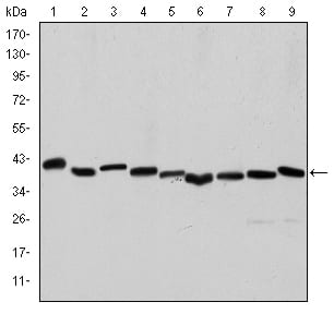
Figure 1: Western blot analysis using ACTA2 mouse mAb against Hela (1), A431 (2), Jurkat (3), K562 (4), HEK293 (5), HepG2 (6), NIH/3T3 (7), PC-12 (8) and Cos7 (9) cell lysate.
Immunohistochemical analysis

Figure 2: Immunohistochemical analysis of paraffin-embedded human duodenum tissues (left) and human esophagus tissues (right) using ACTA2 mouse mAb with DAB staining.
Immunofluorescence analysis

Figure 3: Immunofluorescence analysis of HepG2 cells using ACTA2 mouse mAb (green). Red: Actin filaments have been labeled with Alexa Fluor-555 phalloidin. Blue: DRAQ5 fluorescent DNA dye.
Flow cytometric

Figure 4: Flow cytometric analysis of Hela cells using ACTA2 mouse mAb (green) and negative control (purple).
Elisa

Red: Control Antigen (100ng); Purple: Antigen (10ng); Green: Antigen (50ng); Blue: Antigen (100ng);
For Research Use Only. Not for use in diagnostic procedures.